Publications
Table of Contents (TOC)
Sensing of Vimentin mRNA in 2D and 3D Models of Wounded Skin Using DNA-Coated Gold Nanoparticles
Small, 2018, 14(12), 1703489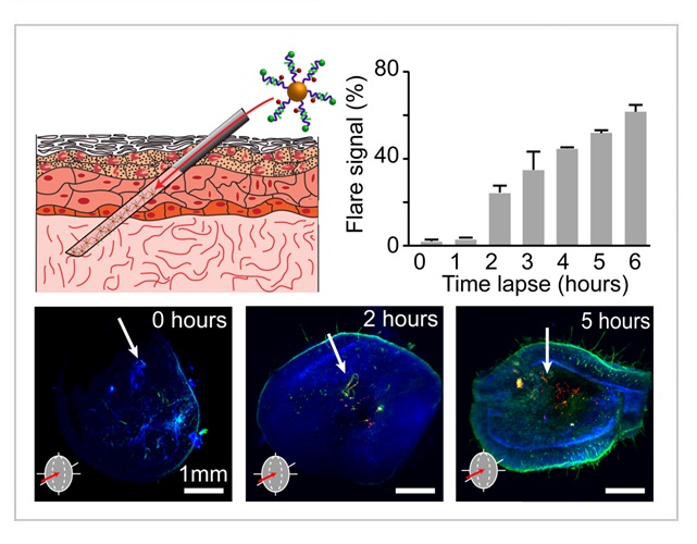
Abstract
Wound healing is a highly complex biological process, which is accompanied by changes in cell phenotype, variations in protein expression, and the production of active biomolecules. Currently, the detection of proteins in cells is done by immunostaining where the proteins in fixed cells are detected by labeled antibodies. However, immunostaining cannot provide information about dynamic processes in living cells, within the whole tissue. Here, an easy method is presented to detect the transition of epithelial to mesenchymal cells during wound healing. The method employs DNA-coated gold nanoparticle fluorescent nanoprobes to sense the production of Vimentin mRNA expressed in mesenchymal cells. Fluorescence microscopy is used to achieve temporal detection of Vimentin mRNA in wounds. 3D light-sheet microscopy is utilized to observe the dynamic expression of Vimentin mRNA spatially around the wounded site in skin tissue. The use of DNA gold nanoprobes to detect mRNA expression during wound healing opens up new possibilities for the study of real-time mechanisms in complex biological processes.
For citation:
Vilela, P.; Heuer-Jungemann, A.; El-Sagheer, H. A.; Brown, T.; Muskens, O. L.; Smyth, N. R.; Kanaras, A. G.
"Sensing of Vimentin mRNA in 2D and 3D Models of Wounded Skin Using DNA-Coated Gold Nanoparticles"
Small, 2018, 14(12), 1703489. DOI: 10.1002/smll.201703489